Limb Reconstruction Surgery Doctor in Mumbai
Dr. Leena Jain, one of the plastic surgeons in Mumbai explains the bony reconstruction procedure and flaps. Bony reconstruction refers to restitution of bone in areas of bony defects resulting usually from injury or from cancer removal surgery. Conventional bone grafting methods effectively treat defects up to 4cms in length if the existing bed enables vascularization of grafts.
In case of longer bone gaps, scarring of surrounding areas and inadequate soft tissue cover ideal option for reconstruction is to use a free vascularised bone graft where bone along with its blood supply is transferred from the lower limb and moved to fill the defect using microsurgery.
Bony reconstruction is required for the following conditions –
Segmental loss of long bones following fractures –
Segmental loss of long bones following fractures occurs due to high-velocity injuries where the bone is fractured in two places and the intervening segment may be crushed at multiple places or maybe bereft of blood supply. This condition is also referred to as a critical segmental defect comprising of a bone void that needs intervention.
Generally, a critical bone defect has a loss of >50% of the circumference or is >2 cm in length. Long bones of the lower limb are essential for weight-bearing while long bones of the upper limb are required for all hand functions. Long bone-bone gaps need to be addressed at the earliest to restore union and function.
Healthy vascularised bone flaps enable union in such cases by bringing vascularised tissue at the ends to be united.
Non-union of long and short bones –
Nonunion is when broken bones do not heal despite suitable surgical and nonsurgical treatment, especially within the initial three months and until nine months after injury. Nonunion is a complex condition wherein comorbidities and detailed evaluation for its resolution are necessary to ascertain the slack in the bony union.
Non-unions can occur in small bones of the hand like finger phalanges as well as long bones like tibia and humerus. Vascularised bone enables a rapid and timely union.
CONTACT US FOR MORE INFORMATION OR BOOK AN APPOINTMENT
Long-standing osteomyelitis
 Osteomyelitis refers to infection of the bone. Osteomyelitis can affect any bone. Chronic osteomyelitis if not treated aggressively can become a long-standing problem severely affecting function. The treatment involves thorough debridement of all infected and dead bone pieces, the release of all surrounding scar tissues, resolution of edema and inflammation, and antibiotic treatment.
Repeated debridements are required at times. Reconstruction of osteomyelitis involves, bringing in vascularised bone and healthy soft tissue with it to reconstruct the bony and soft tissue components.
Avascular necrosis of scaphoid bone
It is a complicated common condition in which the proximal portion of the scaphoid bone does not receive blood supply, and will eventually die. As a result, scaphoidmay collapse, and the patient develops arthritis. Scaphoid fractures are prone for going into non-union and hence replacement with small vascularised bone graft is necessary.
Jaw reconstruction following cancer
Bony reconstruction in the jaw is performed after a mandibulectomy ( jaw removal procedure in case of a tumour in the jaw). The jaw is rebuilt using a bone from a donor site, usually from the fibula l bone of the leg. The fibula is shaped to reform the segment of the mandible removed. The vessels of the fibula are then anastomosed with blood vessels in the neck to re-establish its circulation.
Sites from which bone flaps can be taken:
- Fibula, a bone in the leg, is a flap of choice for most defects requiring a bony restitution.
- Femoral condyle flap used for small bones defects, for treating small non unions.
- Iliac crest taken from the waist – is also used for many defects.
The fibular flap surgery procedure In this surgery, the surgeon removes a bone from the leg, i.e., fibula. The fibula is on the outer side of the leg from the knee to the ankle. Along with the bone, its blood vessels are taken. The flap is attached to the required area of the bone gap and fixed securely with plates and screws.
Blood vessels of fibula are joined to the blood vessels in the neck under the microscope. Once the flap vascularity is restored, healthy bone of the flap brings about the union. Fibular flap surgery is, usually performed under general anesthesia. The patient is required to stay in the hospital for a week
Then, the patient can be made to walk on the operated leg using a non-weight bearing method. It takes nearly three to four months for the union to occur while almost ten months for a solid union.
As a Reconstructive and Hand surgeon in Mumbai performs bony reconstruction procedures using vascularized bone in the following cases –
- Reconstruction of mandibular defects
- Salvage reconstruction of temporomandibular joint
- Reconstruction of bones in the upper limb – humerus, radius, ulna, metacarpal, and phalangeal bones
- Reconstruction of bones in the lower limb – femur, tibia, foot
- Treatment for scaphoid non-union.
CONTACT US FOR MORE INFORMATION OR BOOK AN APPOINTMENT
Medial femoral Condyle flap for bony reconstruction
The medial femoral condyle flap is very useful in treating complex bone defects that require vascularized osseus reconstruction. Also, it provides a large portion of corticocancellous bone that is useful in the reconstruction of the mandible, upper and lower extremities. The descending genicular artery, a branch of the superficial femoral artery, or the medial superior geniculate artery are used to supply blood to the flap.
The medial femoral condyle free flap (MFCF) is a reliable option for treating
- Upper and lower extremity nonunions due to poor bone vascularity
- Reconstructive procedures associated with bone loss
- Avascular necrosis conditions
The main advantage of medial femoral condyle free flap is that it increases bone density, enables primary ossification, and enables healthy blood flow to the area.
The surgeon harvests the medial femoral condyle free flap (MFCF) from –
- The fibula
- The radius
- The scapula
- The iliac crest
- The medial aspect of the knee
Recovery after the bony reconstruction procedure
Generally, recovery after the bony reconstruction procedure takes two weeks to three months. The recovery also depends on the size of the injury as well as the bone graft. The bone graft alone takes three months to heal.
Restrictions
- No extreme exercise for at least 6 months. Seek the advice of a physical therapist before exercising.
- Maintain cleanliness in the bone graft area and ensure it is always dry. Seek special instructions relating to bathing and showering
- No smoking during the recovery period. Smoking slows the recovery procedure. The graft will also fail due to the smoking. Use of nicotine patches is also not advisable.
Outcome of the bony reconstruction
Most bony reconstructions are successful and the bone defects are healed. There is a very minimal rate of rejection of bone grafts.
Dr. Leena Jain, a famous hand surgeon in Mumbai puts to rest most fears relating to failing of bony reconstruction procedures. However, she highlights the need to research and choose an expert and qualified plastic surgeon to perform such challenging procedures.
|
CONTACT US FOR MORE INFORMATION OR BOOK AN APPOINTMENT
Recent Blogs
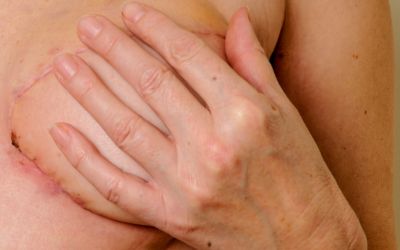
Breast Reconstruction following Breast Conservation Surgery
The journey of breast cancer treatment is full of decisions like the need for mastectomy or breast...
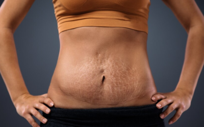
Tummy Tuck Scar After 3 Years
Tummy tuck surgery is a transformative procedure that reshapes the abdomen for a firmer, smoother...

Arm Lift Scar After 1 Year
A year after an arm lift, you reach an important point in seeing how your arm lift scars are...