Happy Clients

Card image cap

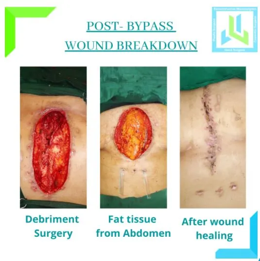
Post- Bypass Wound Breakdown

Heart Bypass surgery is a lifesaving procedure done following symptoms like a heart attack when it is diagnosed the blood supply to different parts of the heart is reduced due to the narrowing of blood vessels supplying blood to the heart.

For bypass surgery, the chest is opened in the midline and surgery is performed. Following this surgery, the wound may take a long time to heal, especially in women due to the weight of breasts stretching the suture line. In some patients, the wound does not heal and opens up exposing the underlying bone- sternum and other tissues. 


These wounds have the potential to become infected, hence, need to be treated early and diligently so as to prevent infection and promote healing. Such wounds are called sternal dehiscence or opening up of the sternal wound.


These patients are then referred to the Plastic Surgeon for wound management. The wound is managed in two or three stages. To begin with, the wound is evaluated for its depth, quantum and type of infection and presence of non-living tissues. A CT scan is performed routinely to assess all these. 


The first stage consists of cleaning up the wound and is called Debridement; following debridement the wound is closed with a special type of dressing called Negative pressure wound dressing wherein a vacuum is created to facilitate better healing.


As the wounding quality improves in 5-10 days, the patient is then taken up for definitive surgery. Most often than not, flap surgery is done to close the wound; wherein healthy tissue is taken from elsewhere in the body and used to fill the cavity of the wound and close it completely. This facilitates complete closure of the wound and optimal healing.

debriment surgery
fat tissue from abdomen


Case Study on Post Bypass Wound Breakdown:

Mrs A, aged 65years, presented to the Plastic Surgery OPD, two months after her coronary bypass surgery (CABG). Her complaints were an open wound with discharge and pain. She said, the suture line after bypass surgery appeared to be healing during the first week, but subsequently she noticed the wound open up in the lower end discharging some fluid and blood.


The upper end apparently continued to heal but the lower end opening seemed to go deeper towards the bone.

We counselled her for the need for a two-stage surgery to which she agreed.


The first stage was debridement – cleaning up the wound and removing all necrotic and unhealthy tissue. The second stage was covering the wound with a healthy flap taken from the abdomen. The wound then went on to heal uneventfully.


As the wounding quality improves in 5-10 days, the patient is then taken up for definitive surgery. Most often than not, flap surgery is done to close the wound; wherein healthy tissue is taken from elsewhere in the body and used to fill the cavity of the wound and close it completely. This facilitates complete closure of the wound and optimal healing.


Post-surgery 2 years and she is doing fine.

After Wound Healing


Card image cap

Forehead Swellings

This middle-aged woman presented with the twin swellings that were evident in her forehead. They were not painful, felt hard and were slowly increasing in size such that they were conspicuous in every photograph!

Once she was counselled about surgery, she readily agreed for the same.

Very commonly people have one but often two swellings on the forehead, usually females. They are bony hard swellings arising from the forehead bone and called Osteomas


They are small in size like marble, hard and remain more or less the same sized. Commonly starting in mid- ’30s, they are painless and cause no other issues except that they are quite obvious on the forehead and stand out in every photograph!


No chance of recurrence!!!


Yes, one can get rid of these swellings by Surgery! It's a simple daycare surgery that can be done under local or general anaesthesia, there will be NO SCAR on the forehead as it will be concealed in the hairline. The excess bone is shaved off the underlying forehead bone.


Her photographs now show a bump-free and scar-free forehead!


BEFORE
AFTER


Card image cap

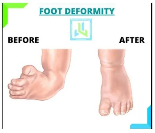
Foot Deformity

This young patient was brought in at the age of 18 years with foot deformity arising from a scar contracture across the top of his foot and toes. Scar was the result of an injury in his childhood which was left to heal by itself without any surgical intervention.

As he continued to grow, his foot deformity worsened as the growth of the bones was hampered by the skin scar. This caused clawing of his toes and altered his gait too. He then came to improve the foot appearance and function to be able to walk normally.


Surgery consisted of release of all scar tissue completely and resurfacing the defect with a flap. Flap was taken from the thigh and using microsurgery the flap was inset into the defect. His foot was maintained in the corrected position using rods, which were removed after 6 weeks.


He was allowed to start walking after about 8-10 days.


With time the flap settled quite beautifully flush with the rest of the normal skin.


Card image cap

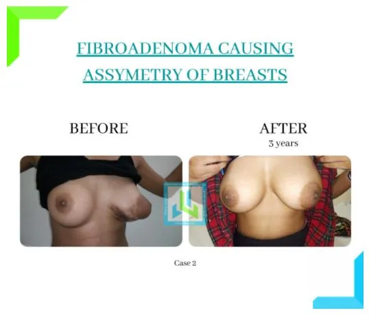
Fibroadenoma

This young 14 years old girl presented with a lump in her left breast of one-year duration. She noticed it steadily in size. It had caused significant stretching of the areola on the affected side. It was a matter in concern as it grew rapidly to the size 10*8cms and caused a disproportionate increase in the size of the breast and the Areola.

She was then evaluated with an MRI scan to see the extent of the tumor. It was found to have two lobes and was extending deep into the breast. Her surgery was planned under General Anaesthesia.


The tumour was removed through a cut along the lower border of the stretched areola so as to leave no scars on her skin. Her breast tissue was then apposed to fill in the gap left after tumor removal. She had an uneventful recovery. Her biopsy confirmed the tumor to be a fibroadenoma.


As she grew up, her breasts grew with acceptable symmetry of the breasts and the areolae, with no scar visible. This young girl was quite satisfied with the overall aesthetics of the breast.


Timely surgery is essential to ensure that breast development is not affected because of a giant fibroadenoma.

BEFORE
AFTER
Card image cap

Hidradenitis Suppurativa

A young male patient working abroad came to us with complaints of Hidradenitis. He suffered from the complication and experienced the following symptoms for these three years recurrent abscesses, discharges, sinuses and scars.

He was frightened about his appearance and social embarrassment. As a result, he avoided many social gatherings. He tried different types of therapies and waited for this long. Finally, he decided to reach us, but it took him three years.


Hidradenitis is a skin disorder in which tiny, painful lumps grow underneath the skin. The tumours may rupture, or tunnels may develop underneath the skin. The armpits, buttocks, groin and breasts are the most common locations where the skin rubs together.


Detailed description

He sought several advanced therapies like monoclonal antibodies and many other prescription drugs, but they all went in vain. After counselling him about the need for surgery as a treatment that would offer a complete remission, he agreed pretty readily.


We removed all the infected, involved, and scarred tissues in a single stage and restored the armpit dimensions to normal. The flap was taken from the chest wall side and turned upwards to resurface the armpit defect.


Healthy tissue replaced in the armpit was never affected by Hidradenitis ever, and it gave a pleasing contour and colour match.


BEFORE
AFTER
Card image cap

Wrist Amputation

A 4-year-old girl was playing outside her house when she heard a recorded voice from the elevator calling for the door to be closed. Her right hand got trapped in the door when she attempted to close it. 

The child’s wrist tore offas the lift climbed to the fourth floor. In a 12-hour surgery in the middle of the night, a team of Plastic Surgery and Hand Surgeon at Lilavati hospital successfully reattached her wrist.


She had had an unfavourable amputation where all vital structures such as nerves, blood vessels, and muscles were pulled from her wrist separating it from the rest of her forearm.


The surgery was difficult as it required fixing the bone, joining tendons that drive the thumb and fingertips, joining major nerves that bring sensations from the hand and send signals from the brain to the various muscles in the hand and joining major arteries and veins to restore blood flow to the hand.


For the first seven days, the reattached hand was fine, but on the eighth day, it became cold and blue. Doctors were notified right away, and the child was taken back in for surgery. The veins on the back of her hand were discovered to be blocked. 


They were seriously injured at the time of the accident, and a clot had formed within them, blocking blood flow. Additional veins from her leg were used to patch the veins once more. She then underwent another major microsurgical procedure, this time transferring muscle from her thigh to her wrist to cover the region of skin and soft tissue loss.


She was then discharged with a healthy right hand and continues to be on regular follow up.

Here is the Newspaper publication of the above case – Midday


BEFORE
AFTER
Card image cap

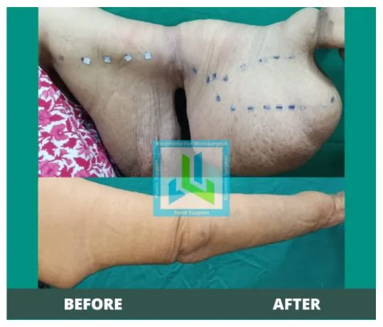
Lymphedema of Upper Limb

This cheerful lady, in her sixties, presented with swelling of her left upper limb since last 10 years after she underwent surgery for left breast cancer (in 2010). 

The swelling kept increasing with time, while she went on modifying her right-hand dexterity to go about her routine household work without ever complaining. Unaware of the surgical options for this, it reached an extent of growing to the size of about over 4 kgs.


Her family came in a quest for a possible long term solution. Her patient disposition made it easy for her to understand the compliance required post-surgery.


Surgery

As it was in a late-stage, we debulked the entire upper limb with a combination of liposuction and excision. The entire tissue was removed in a single stage in her case, with a good circumferential reduction.


With the help of regular exercises, compression garment and self-care, she is quite content, uncompromisingly going about her household activities.


Book an Appointment

Enter details,our team would approach to help you as soon as possible.

Phone icon
Call Now